




Product Description
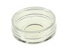
Image live cells at high resolution without moving them off their growth surface. These 35 mm dishes have a thin borosilicate glass bottom, so cells grown in the dish can go straight onto confocal, fluorescence, DIC or phase contrast microscopes.
This listing is for NEST catalog number 801001: 35 mm Glass Bottom Cell Culture Dish, 20 mm Glass Well, TC-Treated, Sterile, Case of 200 (20 Packs x 10). The glass is 0.16–0.19 mm thick, has low autofluorescence and is treated for adherent cell attachment. It is bonded to a high-clarity USP Class VI polystyrene dish with a non-cytotoxic, medical-grade adhesive. Dishes are molded and packed in an ISO 7 cleanroom in a cGMP, ISO 13485 and ISO 9001 facility, E-beam sterilized to SAL 10⁻⁶, and lot tested pyrogen-free. Each dish comes with a 15 mm or 20 mm glass well. Order it from LIGHT LABS alongside the rest of your NEST plastics and consumables.
Key Features
- Thin Glass Bottom – 0.16–0.19 mm borosilicate glass for high numerical aperture objectives
- Low Autofluorescence – Clear background for fluorescence and confocal imaging
- TC-Treated Glass – Surface prepared for adherent cell attachment
- Two Glass Well Sizes – 15 mm or 20 mm imaging area in a standard 35 mm dish
- Non-Cytotoxic Bonding – Medical-grade adhesive secures the glass to the dish
Specifications
| Catalog Number | 801001 |
| Brand | NEST Scientific |
| Description | 35 mm Glass Bottom Cell Culture Dish, 20 mm Glass Well, TC-Treated, Sterile |
| Sold As | Case of 200 (20 Packs x 10) |
| Glass Diameter | 20 mm |
| Units per Pack | 10 |
| Units per Case | 200 |
| Dish Diameter | 35 mm |
| Dish Material | USP Class VI polystyrene |
| Glass | Hydrolytic borosilicate, 0.16–0.19 mm thick |
| Surface Treatment | TC-treated |
| Adhesive | Non-cytotoxic medical grade |
| Sterilization | E-beam, SAL 10⁻⁶ |
| Certified Free Of | Pyrogens (USP <85>) |
| Manufacturing | ISO 7 cleanroom; cGMP, ISO 13485 and ISO 9001 facility |
Available Options
Applications
- Live cell imaging
- Confocal microscopy
- Fluorescence microscopy
- Differential interference contrast microscopy
- Fluorescence in situ hybridization (FISH)
- Phase contrast microscopy


